What does corneal topography measure?
Of all the tests in a refractive surgery assessment, corneal topography is probably the most important. It shows whether your cornea is regular and thick enough for laser treatment. An abnormal map alone can rule out LASIK or even any corneal surgery. Here is what the test measures, how to read a map and what your surgeon is looking for.

What the tests measure and how
The cornea is a transparent dome about half a millimetre thick at the centre. Your surgeon looks at three features.
- Curvature at each point, which determines focusing power and astigmatism.
- The shape of the front and back surfaces. Early keratoconus often changes the shape of the back surface first.
- Thickness at each point, to assess how much tissue can be removed without weakening the cornea.
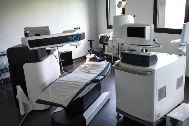
The Centre médical des Aravis uses two complementary technologies.
| Device | How it works | What it shows |
|---|---|---|
| CA-800 corneal topographer | Analyses the reflection of illuminated rings (a Placido disc) on the surface of the eye | Detailed front-surface curvature, pupil measurements and analysis of the tear film and eyelid glands |
| ANTERION tomographer | Cross-sectional imaging using swept-source OCT | Shape of both surfaces, a thickness map, anterior chamber depth and measurements for implants |
The test is quick and the device does not touch your eye. You look at a light for a few seconds while it takes the images. The centre’s equipment, on annecy-ophtalmo.fr
How to read a map
Maps are colour coded. Warm colours (red and orange) indicate steeper or higher areas, while cool colours (blue and green) indicate flatter areas. The surgeon examines several maps and measurements.
- The curvature map. A symmetrical, vertical bow-tie shape indicates regular astigmatism. An asymmetrical or tilted bow tie, or a red area below the centre, can suggest an irregular cornea.
- Front and back elevation maps compare the cornea with a reference shape and show bulges too subtle to see on the curvature map.
- The thickness map (pachymetry) shows the thinnest point and how the cornea thickens towards the edges.
- Automatically calculated indices help classify the cornea as normal, suspicious or abnormal.
Your surgeon reviews the results together and compares your two eyes. They may also compare two examinations before reaching a conclusion.
Keratoconus and the risk of ectasia after surgery
Keratoconus is a condition in which the cornea gradually thins and bulges, usually from adolescence. It is more common than once thought. A Dutch study from 2017 estimated that it affects around one person in 375. Early keratoconus sometimes shows up only as ordinary-looking astigmatism on a prescription. What the axis means on your glasses prescription
Laser surgery on a vulnerable cornea can trigger ectasia. The cornea, weakened by the removal of tissue, changes shape much like it does in keratoconus, causing a loss of vision that is hard to correct. Ectasia is rare and topography is the main way to prevent it. Known risk factors include an abnormal map, too little residual tissue under the treated area, young age and a thin cornea before surgery.
If your topography is suspicious, the surgeon may repeat the test after a few months, rule out LASIK in favour of another option such as a phakic implant, or monitor the cornea. For progressive keratoconus, cross-linking can strengthen the cornea and slow the disease. Keratoconus, on annecy-ophtalmo.fr · Cross-linking
What can affect the test results?
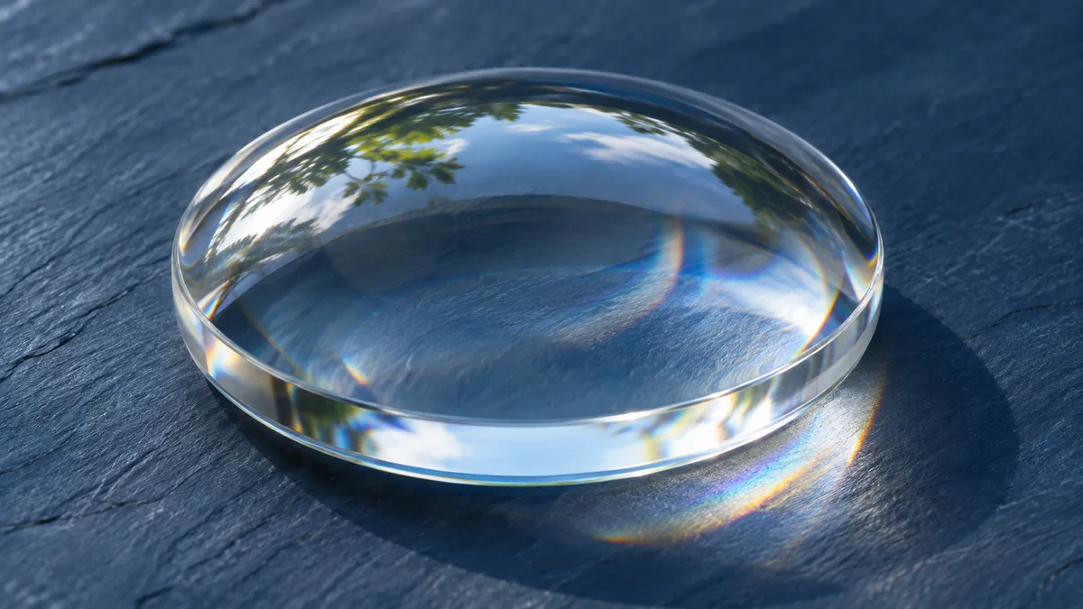
- Contact lenses temporarily change the cornea’s shape. Stop wearing soft lenses at least one week before the test and rigid lenses at least three weeks before. Contact lenses and your assessment
- An unstable tear film distorts the reflected patterns. Dry eye may need treatment before measurements are repeated.
- Rubbing your eyes, which is common with allergies, weakens the cornea and changes its shape. Tell your surgeon if you do this.
In summary. Topography measures corneal curvature, the shape of both surfaces and thickness. It checks for early keratoconus, the main risk factor for ectasia after laser surgery. A suspicious map can rule out LASIK in favour of another option. Stopping contact lens wear in good time is essential for a reliable measurement.
Frequently asked questions
Does an abnormal topography mean I have keratoconus?
Contact lenses or dry eye can distort a map. Your surgeon compares the test results and may repeat the measurement.
Is topography repeated after surgery?
Yes, it is often part of follow-up. It helps check that the treatment was centred and allows the surgeon to compare the cornea with its original shape.
Can I see my results?
Yes. Ask for a copy of the maps. They are a reference for future examinations.
To have your cornea checked, check your eligibility and see what to bring to the assessment.
Sources
Related reading
All news
News
When your myopia prescription changes
Before refractive surgery, the first question is whether your myopia is stable. A laser corrects the myopia measured on the day of…
22 April 2026

News
Who assesses fitness for jobs with eyesight standards?
For some jobs, eyesight is part of the fitness requirements. Pilots, air traffic controllers, professional drivers, military personnel, firefighters and police officers…
27 August 2026

News
Screen eye strain: what to tell your surgeon
Stinging eyes at the end of the day, blurred vision on a spreadsheet and headaches after a video call are typical of…
22 July 2026
Have a question about your situation?
An assessment tells you what is and isn’t possible and allows you to get a quote.